REDOX Rx 2: BIOHACKING YOUR MRI
READERS SUMMARY:
1. HOW CAN AN IMAGE OF PROTONS TELL US A TON ABOUT YOUR REDOX POTENTIAL IN YOUR CELLS?
Today’s blog is stimulated by a challenge I received during the epic 12.5 hour Q & A that occurred after a talk I gave in Pasadena in September. During the period an older radiologist challenged my statement that there was no way that I could look at a MRI image of one of my patients and deduce anything about their diseases, physiology, anatomy, or redox potential. The topic came up because I casually mentioned some of my MRI bio-hacking tricks of the trade to diagnose osteopenia and osteoporosis to another anesthesiologist who was in our group. I explained to both physicians the more data we can pull out tests already done on patients the better prepared we can be for things we may face in our treatment plans. The anesthesiologist seemed to get that insight, because he understands in surgery we need to prevent problems before they occur by any means possible to gain extraordinary outcomes. I wish I could say the same for our radiologist colleague.
The radiologist challenged me with his dogma, and not facts. I tried to be gracious to him, but it was clear to me, and those in attendance, that I could not explain the physics of MRI imaging to him in a short period of time without boring the crowd. I invited him to come my forum or speak with me off line, but neither one happened. I was in teaching mode but this fellow was more interested in pushing the conventional wisdom agenda that I usually encounter in a hospital. I mentioned to one of the young ladies who was in the crowd, who was a medical student, that I was going to revisit this situation in a blog. I felt it was too important to let fester. If we do not strive for excellence we will wind up with mediocre, and that is just not good enough for patients.
To be able to see or appreciate something, you must first know or believe it exists, or at least ,not have preconceived ideas that it just can’t happen. Here is where the rubber meets the road in this teaching point.
I assured him that it was not only possible but based upon a scale of science he or I did not learn in medical school or residency. I believe I infuriated him when I told him that I believed he was a sucker to his beliefs that were given to him by his mentors in radiology. I know it was an “ego blow” to have a surgeon tell him, that something he was supposed to be an “expert in” reading had a lot more depth of information about matter and energy than he could fathom.
So today’s blog is about going back to the fundamental science he just could not accept.
How much do you know about proton spin?
In 1952, the Noble Prize was given to Felix Bloch and Edward Mills who introduced the concept of nuclear magnetic resonance (NMR) to chemistry and physics in 1946. The quantum idea buried at the heart of the NMR is that one can voluntarily “force” the alteration of atomic orientation of atoms by altering the orientation of the magnetic moment of all atomic nuclei. We can do this by entering into a resonance with them. A resonance is a frequency, oscillation or vibration. When we enter a resonance with atomic nuclei, we can modify their overall magnetization by giving them a specific energy in the form of a radio wave at a very precise frequency to disrupt that resonance and measure the effects.
It is important to point out that not all atomic nuclei are capable of being magnetized. Those made up of an even number of protons and neutrons do not have a magnetic moment. When the number of both is even, magnetic effects are nullified within the atomic nucleus. I turns out that life uses this to create stability and growth. This is why H+, C14 and O16 are key atoms of proteins in use that are designed to be long lasting and stable in our magnetic field on earth. This also happens to be why radiologists have fallen in love with the H+ proton for image generation for MRI’s. Hydrogen forms 2/3 of water molecules, is abundant in us, and makes up 99% of the molecules by number in our body. Water happens to make up 80% of the brain’s mass. We have 30 millions of billions of water molecules in us! So this makes protons vitally important to the mitochondriac.
Think mitotic spindles in cells
Water, however has some interesting properties that our radiologist friend might not have known. Water is what hides quantum mechanisms from biology. Water is a cell’s natural Faraday cage. It also is special inside cells because of how its made. We live in world of the infinitely small where quantum physics dominates all biologic effects inside of mitochondria. And that world is quite different from the world we observe. The job of a quantum clinician is to teach a patient how to see the things with their mind that their eyes cannot observe and MRI’s happens to be one of my major teaching tools in my clinic.
In a magnetic field of 1.5 tesla, 1 tesla represents 20,000 times the strength of the magnetic field in New Orleans. Protons from water can line up in the direction of the magnetic field, or in an opposing orientation. The direction of their “lean” depends tremendously on the intensity of the magnetic field they sense , which is determined by the difference in the number of protons in these two orientations. It turns out the difference of a 1.5 tesla and 1.0 tesla magnetic field is seen in only 50 protons per 1 billion. At 3 tesla’s it rises to 100 protons per billion. Since we have 30 million billion water molecules in us, it begins to make a huge difference in what the observable magnetic moment is. So it should make sense now why higher Tesla MRI’s give better images. They are able to sense more hydrogen protons. H+ is just a single proton with its electron stripped off. This is the dominate form of hydrogen in the mitochondrial matrix where the most critical energy pathways are located. 30% of humans cells are made of mitochondria. This makes MRI a very sensitive tool for assessing mitochondrial wellness. This ratio of H+ is what determines the data on an image in a MRI. This is why MRI is a study of the atomic physics of water. Water is how we decipher environmental signals. It is not an homogenous fluid as most believe; it is an ionic plasma that imprints energy and information from the atoms around it.
This is why I decipher a lot from MRI and why our radiology critic thinks we can’t see osteoporosis on MRI’s. Hydrogen isotopes can alter the atoms around it differently than H+ will. That alteration also effects the proton signal in MRI images. Protons are like “shadows”, in this sense. When I see an MRI, I am reading the “shadow cast” of the hydrogen proton. MRI images are not real images, they are virtual sections emanting from an ephermeral magnetizaion of water in the tissue being imaged. My ‘radiology friend’ could not comprehend this because of what he was taught classically in residency was devoid of physics. When you understand the physics of NMR and hydrogen protons, you begin too see why I said what I did, and why he was adamant he could not see disease states in a MRI alone. You can’t see a shadow you don’t learn that exists, can you? MRI’s are a GPS for a neurosurgeon because we use it to navigate while a radiologist only reads the films and never correlates it with the patient’s real anatomy. In this way, neurosurgeons develop a keen sense of what is really possible with the magnetic shadow cast of the MRI, if they are paying attention to the physics that generated the image.
What else did the radiologist fail to grasp? NMR or MRI is useful because nuclei of atoms inform us about their local environments on an atomic scale. He failed to understand that hydrogen is not just found in water but it is found in many biologic molecules life uses, such as amino acids, proteins, and nucleic acids. He also forgot basic organic chemistry, it appears. NMR is used to decipher what atoms are in a specimen, whether it is in a test tube or a MRI gantry. The irony here, is every MD in the US has to take organic chemistry to get into to medical school. As part of our training we were required to purify certain organic chemicals by experiment, and then use NMR to see how good a job we did. NMR = MRI. The science is identical but the name is different because when MRI was invented scientists and clinicians were were worried no one would have a body NMR scan because it contained the word nuclear. So they made up a new name for the scan, called magnetic resonance imaging. This is how I decipher the redox potential so well from an MRI image. I mentioned his ability in the Redox Rx blog.
Since the environment “upsets” (think metastability) the local magnetic environment in the nuclei of atoms creating a cell, this change is translated by small variations in the frequency of the resonance of the waves that are reemitted by the atomic nuclei in question. By careful analysis of these waves, we can decipher their frequencies precisely. This allows us to deduce the exact molecules in which these nuclei are found, in what quantity, and even in what part of the molecule they are in! What else did he miss?
The EMF Part
Since the frequency of the resonance of waves emitted by the atomic nuclei is wholly dependent upon the magnetic field it is found in, we can see the images of the actions of these nuclei when the field is progressively altered. This is how gradient images are mapped to make image we neurosurgeons use every day. If you’re reading between the lines, you can also see you can tell when someone lives in a very altered electromagnetic field too!!!. You will see marked abnormalities in their tissues that are not explained by their age or disease status. This was my big clue why osteopenia epidemic was linked linked to our non native EMF exposures. Non native EMF fields also cause alterations to the magnetic field and those changes are transmitted to atoms in us!!!! The coils in an MRI machine are used to generate different magnetic fields to make pictures using helium to cool the magnets to improve their strength. In disease states, people also face different magnetic fields in their environments and from a slower spinning ATPase rotating head, but few radiologists realize this, but the data is always in the images. Radiologist dictate what they are taught to see and not they do not mention the things they were not taught. The coils in you (nucleic acids), electric circuits you face now daily like ipods, laptops and TV’s generate the non native magnetic fields to slow spinning of your ATPase to cause diseases in you because mitochondrial redox power is lost. Native magnetic field signals are blocked and can be interfered with by non native EMF signal’s made by man.
For those of you were there in Pasadena in 2014, you can see why I scoffed and just moved on from his questions during that Q & A. He fundamentally had no clue how MRI images were generated using the laws of physics, so he had no educational basis to tell me I could not do what I do with MRI’s in the clinic. That is how dogma blocks us from progress in medicine. I believe we can teach clinicians this interpretative science to allow them to become expert in determining the redox potential of their patients tissues by looking more critically at their MRI images.

Often these images are already done for other reasons, which makes them more cost effective. Moreover, we can fact check my interpretations and theories if we use MRI spectroscopy. This test will allow us to reconstruct 3D atomic structure of molecules that we can harvest via percutaneous procedures done for other reasons. MRI spectroscopy extends our insight by also allowing us to develop a temporal dynamic for the molecule in question. Richard Ernest won the Nobel Prize in chemistry for deducing this fancy quantum mechanism out, but it seems few radiologists are learning to use it as I have. The science is there, but you must taken the science already discovered and look at it a different way, to consider something in a new way no one has thought about before. This is what an innovator does.
It does not diminish the person who does this…….it elevates mankind’s knowledge base of what is possible. This is why I was so irate with this “supposed expert” in Pasadena. I promised those who were present I would post a blog about this aspect of the Q & A, and now you have it. MRI’s have a ton of atomic data in them, if you know what to look for. I look for alterations in quantum spin of protons.
Well, I am in Asia and jet lagged after 28 hours of travel. I will be giving several talks to hundreds of interested physicians so you can expect another blog down the pike about this experience. I am appreciative of the chance to spread the message of how we can use physics to change our biology and our life.
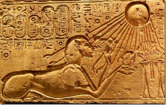
Photonics over Electronics is an ancient meme that modern man’s narrative for living has ignored at our peril.