

READERS SUMMARY:
1. HOW DOES MYELIN DO WHAT IT DOES?
2. DO WE REALLY UNDERSTAND HOW MYELIN AND WATER CHANNELS WORK?
3. WHY DOES MULTIPLE SCLEROSIS REALLY HAPPEN?
4. IS MULTIPLE SCLEROSIS DUE TO A SHORT CIRCUITING OF MITOCHONDRIA BY EXCESS LIGHT?
Do you know the story of the Japanese AMA? If not have a look here. The Japanese AMA never have had a reported case of MS. Multiple sclerosis (MS) in Asian populations is characterized by the selective and severe involvement of the optic nerve and spinal cord as well as low prevalence rates. The Japanese eat a lot of DHA naturally. Might this be why they have a different form of MS than the rest of the world? Why is its incidence and prevalence exploding since 1960? Japanese people born after modernization in the 1960’s, the ratio of conventional to opticospinal MS has increased rapidly. Technology has also exploded in Japan since 1960. Let us explore the links.
Multiple sclerosis (MS), an inflammatory demyelinating disease, is a major cause of neurological disability in young adults in the developed world. Although the progressive neurological disability that most patients with MS eventually experience results from axonal degeneration, little is known about the mechanisms of axonal injury in MS. I believe the reason for this is because no one is looking at the quantum scale to see how this disease forms. Today, accumulating evidence suggests that the increased energy demand of impulse conduction along excitable demyelinated axons and reduced axonal ATP production induce a chronic state of virtual hypoxia in chronically demyelinated axons. Hypoxia is tied to a lack of ATP production and low O2 tensions. These are associated with a loss of heat from the inner mitochondrial membrane. Heat happens to be a form of electromagnetic spectrum of light. Heat is a associated with proton flows in mitochondria.
ATP production is decreased when humans naturally uncouple oxidative phosphorylation. It turns out, MS patients are chronically uncoupled by several environmental stressors that few are tying to the cause of this disease. This means MS patients are losing proton flows constantly. I believe the results Pollack has found in water is a very significant part of the MS story being missed; it is much easier for H+ to dissociate from the water molecule than for the water molecule to lose an electron.
Uncoupling is normally done in the non pathologic process by cold environments allowing mitochondria to release heat. People with MS are very temperature sensitive both at cold temperatures and hot temperatures. This is because they all have abnormal T3 function and they cannot properly use UCP1 or 3. When you are temperature insensitive it implies you have lost the ability to control uncoupling for some reason. Uncoupling allows for loss of heat. Heat is a form of light called infra-red light and is tied to proton flows. It has been experimentally shown by Fritz Albert Popp that Multiple Sclerosis patients absorb excessive amounts of light because they are losing excessive amounts of heat and protons from their mitochondria. In essence, MS patients are drowning in spectral deficient light and this causes then to leak protons constantly from their mitochondria and this lowers the exclusion zone in cells and as a secondary effect the interior of the cell become less negative.
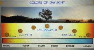
Remember light covers many spectral bands and frequencies. MS patients seem to be blue light toxic from artificial sources. Blue light is associated with long light cycles, carbohydrates, and modern technology gadgets which have high color temperatures.
It turns out Popp experiments revealed that nucleic acids, ROS, and RNS release light within cells when our cells are under any stress. In the 1970’s, Dr. Fritz Albert Popp coined the term “Biophotons”, for the ultra weak photonic emissions given off by cells during reactions. This light is very weak – typically several million times as faint as the light from a firefly. He was able to confirm that living cells emit small bursts of light normally. He determined that cells do not just radiate light, they can also absorb light. Popp found in his experiments that healthy cells tends to release very few photons, maybe only ten per minute. Cells that are not healthy leak a lot of light, according to Popp. He also found these photons were not easy to collect, even in a pitch-black lab. In the 1980’s his job got easier in collecting this light emission. Researchers developed light detectors called photomultipliers and researchers found these emissions coming out of everything from plant seeds to fruit flies and to all animals.
The number of photons emitted seemed to be linked to the organism’s position on the evolutionary scale — the more complex the organism, the fewer photons were emitted.
•Humans emit only 10 photons/cm2/sec at a wavelength of 200-800 nm.
• Rudimentary animals and plants tended to emit 100 photons/cm2/sec at a wavelength of 200-800 nm, corresponding to a very-high-frequency EM wave well within the visible range
• Microbes like bacteria emit over 1000 photons/cm2/sec , and drastically increase their ability to emit biophotons when in growth phase at a low (107) concentration. You might remember that mitochondria are related to bacterial microbes from an evolutionary standpoint. This is why mitochondria leak certain frequencies of light that can effect cytochrome c oxidase and the ATPase. Eukaryotes assimilated a bacteria within its cell and it became a mitochondria 600 million years ago. This process was called endosymbiosis. This means that uncoupling in mitochondria is a vestige back to their bacterial origin. Mitochondria also divide by binary fission and use lateral gene transfers in cells to give tissues an energetic footprint that varies tissue to tissue. Eukaryotes have used their ability to leak light to our advantage in uncoupling in environments outside the tropics. These are the environments MS occurs.
People with MS have poorly functioning mitochondria because of defects in their light environments. These are all releated to spectral deficiency’s that lead to poor energy flows from mitochondria that support the aquaporin 4 water gates in the CNS. In Popp’s experiments unhealthy cells always seem to release more light than healthy ones.
Popp found people with MS were absorbing lots of light while “losing light” more rapidly from their cells.
This data is the key missing piece to understanding MS from a quantum basis, in my opinion.
People continue to believe they need a balanced diet of high quality food. What they really need is a balanced diet of solar light. Solar light is not equivalent to artificial light. Man made light is polarized and spectrally deficient. We not only “eat” sunlight–we can actually digest it and break it into its component frequencies to power specific mitochondrial processes that drive the neural processes that control growth and metabolism.
When mitochondrial energy flows are low it effects other bacterial colonies we harbor. That is mitochondrial oscillation in adjacent tissues and the bacteria in out gut. They begin to simplify. The mode of energy and data transfer can be found in my Time 22 blog.
It should make sense to you, because food is really just a proxy for electrons that sunlight as has excited in the photosynthetic web. Light has always been primordial to life on earth, while food has evolved with growing seasons. It evolved under the power of sunlight we call the quantum yield. Life had to be built from light at its origins. There was no other choice. Even food’s chemical bonds ultimately derive their energy from the sun and their bonds are effected differently by different frequencies in the spectrum of light. This makes man made light a real problem for people with MS.
Sunlight is supposed to provide us a “balanced diet.” Understanding MS fully is beginning to understand how modern artificial light is really highly-unbalanced “junk food” for our mitochondria. Red and infrared light are more like organic “health food” for a mitochondria because of its effect on cytochrome c oxidase and the ATPase. Blue light destroys the voltage in mitochondria and slows electron tunneling speeds to reduce energy efficiency. In fact, Popp has shown that 380-nanometer light is a “superfood” for regeneration. Several researchers have backed up this claim with experiments. The frequency of light in our bodies is critically important. Light can only enter our cytochrome proteins as incident light when it excites electrons of foods which ferry it to our mitochondria after we eat. UV-light is like caffeine or “jet fuel” for the mitochondria and this is why electrons excited by UV light enter at the NAD+/NADH couple ( it can help or hurt depending upon the space within your cells). Gamma rays and x-rays are most always poisons because they excite electrons closer to to the nucleus. Solar light only effects the valence electrons. Radio-waves (nn-EMF) when present in excess, by interfering with magnetic perception, cellular signalling, and mitochondrial electron transport basically cause a “light indigestion” in mitochondria. Cell phone antenna’s all use an RF antenna. Many people put these devices up to the head chronically in the modern world.
“Minding your mitochondria” has much less to do with watching your macronutrient intake than it does with managing your frequency intake!
THE QUANTUM CONNECTION TO LIGHT:
In line with quantum electrodynamics theory, the exclusion zone water appears to be formed by the interaction of light with the water surrounding our mitochondria. This has been experimentally shown by Pollack and many other labs now. The most effective type of light in forming exclusion zone (EZ) water is far infrared/microwave ~3000 nm. The gap between the ionizing potential of 12.56 eV and the excited level of 12.06 eV predicted by Emilio Del Giudice and colleagues is 0.5 eV, equivalent to 2479.7 nm, not too different from ~3000 nm (equivalent to 0.413 eV); although the 3000-nm absorption may have more to do with the rearrangement of coherent domains (CDs) into EZ water rather than charge separation. Here Del Giudice science makes practical application of Pollack’s experiments already completed in our mitochondria. EZ water has been shown to increase in electrical conductivity by several orders of magnitude.
KEY MS POINT: MS patients lack EZ water around their mitochondria and organelle’s because most suffer with high BUN to creatine ratio’s.
When we discuss light many people think light is “visible light” (400nm-700nm) when we mention it. The above paragraphs paints a different picture of the light contained within your mitochondria. The light at cytochrome 1 is UV light (340nm) and the light inside of a mitochondrial matrix is mostly infrared. Infrared light is heat and it can glow red if the intensity is present. In us, the intensity is not great but researchers have shown bio-photons are released from all human organelles. Healthy cells tend to emit a constant phase relationship during the course of time. Unhealthy cells emit a non-constant, diffuse phase relationship during the course of time. This is how we fundamentally uncouple oxidative phosphorylation in mitochondria when we are in cold weather. It is a vestige of the microbial light emission mechanisms I mentioned above. Bacteria normally emit large amounts of IR heat. Our mitochondria is a vestige bacteria. Our mitochondria normally release heat as well when we uncouple oxidative phosphorylation. Uncoupling stops ATP production in mitochondria but electron chain function continues to deliver electrons more slowly to reduce molecular oxygen. The speed of electron transport and tunneling reduces the amount of oxygen made and this leads to chronic pseudo-hypoxia in neurons.
As mentioned in the opening paragraph, MS patients never fully reduce molecular oxygen with electrons because of this chronic uncoupling affect. This creates a state of chronic hypoxia, dehydration, and stress within neurons. People with MS can’t retain their IR heat because they have simultaneously LOST the high electric charges on their cell membranes due to many environmental stressors most of them are unaware of. So are their physicians. Recall that cell membranes lose DHA as “blue light stress” rises. This happens disproportionally within their eyes, skin, and CNS where mitochondria populations are numerous. This is excerabated when blue light is the main driver of the stress. You would be wise to remember skin and brain cells are both derived from embryologic neuroectoderm. This implies that both tissue’s mitochondria are functionally and morphologically related and linked by oscillations. (See Time 22 for the mechanism)
DHA is a natural ligand for retinoid X receptors (RXR= Vitamin A) – obligatory step – stimulates > 107 genes that developed the brain 600 million yrs ago. DHA has the ability to turn light into electric signals. DHA is where optogenetics began in evolution. Everything was fine……..until humans invented fake light and stopped eating seafood.
Nutrients and many other environmental factors have also been found to influence epigenetic programing of our DNA either directly or indirectly via metabolic sensors. Peroxisomal proliferator-activated receptors (PPAR’s), the vitamin D receptors, and the retinoid X receptors (RXR=Vitamin A cycle), and the retinoic acid receptors (RAR) are all examples of the nuclear receptors that interact with the brain cell membranes to control inflammation and metabolism all over our bodies. It turns out that PPAR’s are the receptors that are at the crossroads of where inflammation and metabolism actually cross. This system is destroyed in MS. Most people understand in MS vitamin D levels are quite low. No one can explain why. I think this blog will.
it also turns out that fish based DHA (not a supplement) is also a ligand of retinoid X receptors (RXR), and RAR binds most of the forms of Vitamin A in our bodies. Remember from the Hormone 101 blog to make hormones we need Vitamin A and T3 to be present in good concentrations to convert LDL cholesterol to pregnenolone in order to make our steroids. Pregnenolone then undergoes transformation to the rest of hormone chain in humans, provided cytokine levels are low. That hormone chain involves Vitamin D production in the skin making it an ideal way to tell us what is going on in that engine we call a mitochondria.
This loss of charge in our cell membranes directly affects electron flow on the inner mitochondrial membrane in MS. When ECT transport is slowed down for any reason, humans make more free oxygen radicals, and less O2 and increase their reliance on carbohydrate metabolism. These radicals cause further release of IR light from the mitochondrial matrix. The inner mitochondrial membrane, the protein-to-lipid ratio is 80:20 where the cytochromes and transition metals are located, in contrast to the outer membrane, which has 50:50 mix of protein-to-lipid ratio. The outer membrane is directly connected to the endoplasmic reticulum where protein synthesis or dys-synthesis can occur by affecting tertiary and quanternary bends on proteins. These bends will affect how light can interact with these aquaporin proteins in neurons. The proteins on the outer membrane do not have as many transition metals by design. The released heat affects the exposed transition metals in their cytochrome proteins because they lose their ability to conduct electric signals and tunnel electrons rapidly. Recall that electric resistance rises as temperature increases. Popp found experimentally these free radicals cause light loss in his research. The exposed light interacts with these metals and also causes reactive hypoglycemia as well.
When anything suppresses mitochondrial respiration, it exacerbates hypoglycemia. Here you see where carbs and light affect MS. Very few people in ancestral health seem to realize this basic mitochondrial effect. With time, sympathetic outflows from the PVN will compensate for this loss and cause an increase in blood glucose to offset this reduction in blood glucose. This is why the most common finding of blue light toxicity is adrenal fatigue. If this process becomes chronic it leads to things like MS. This causes people with MS to rely too heavily on glucose metabolism and AMPK pathways to maintain metabolic balance in their mitochondria. This occurs due to calcium efflux from the PVN in the brainstem. This is also why most people with MS are found to have adrenal fatigue when salivary adrenal stress indices are checked by clinicians. Few do because they have no idea what really causes MS. MS is a blue light toxic disease that slowly poisons CNS mitochondria.
Nora Volkow and Abraham Liboff work has shown similar effect’s in their research with other non native EMF’s. Mina Bisell has found the same thing in breast cancer research. I will remind you here that blue light is a form of non native EMF for humans. AMPK up-regulation is also tied to excessive carbohydrate fuels outside of the proper circadian seasons in people with MS. This is NOT GOOD for people with MS on a chronic basis because it exacerbates the chronic hypoxia found in mitochondria that destroys myelin. Popp also found that healthy cells also exhibit a coherence regulation to the Earth’s normal circadian rhythms, while cancerous cells and autoimmune disorders did not. MS is an autoimmune disorder and one that is linked to an environmental mismatch of artificial light that causes alterations in circadian signaling.

Think about how mitochondria work to tunnel electrons normally; when the inner mitochondrial membrane loses its electric charge, it increases the mass in mitochondria by inducing swelling. This redox shift in mitochondria is most commonly seen when circadian signaling is off. This swelling signal is used to activate or deactivate the two recycling programs in mitochondria called autophagy and apoptosis. In both processes, the cell membrane gets less dense and the size increases. When the inner mitochondrial membrane swells it stops the rotating head of the ATPase from spinning. This stops ATP formation. The swelling also slows electron tunneling from complex one to the oxygen. Oxygen has a special magnetic order that is lost MS because of the chronic hypoxia due to poor mitochondrial function. Triplet oxygen (not to be confused with ozone, O3) is the ground state of the O2 molecule in mitochondria. The electron configuration of the molecule has two unpaired electrons occupying two degenerate molecular orbitals. In normal triplet form, O2 molecules are paramagnetic. That is, they form a magnet in the presence of a magnetic field—because of the spin magnetic moments of the unpaired electrons in the molecule, and the negative exchange energy between neighboring O2 molecules.
Recall that the human ATPase as a spinning head. Rotation of the “g shaft” of the ATPase relative to the ring of a and b subunits was demonstrated by H. Noji, R. Yasuda, M.Yoshida & K. Kinoshita. This acts like a windmill that can rotate the power of a magnetic field or from the energy in a photon of IR light. The lack of spinning of the ATPase also reduces the amount of oxygen reduced and produced in mitochondria; this results in a lower powered magnetic field effect in every mitochondria of MS patients. When this effect is lost, swelling occurs more easily because the mass of the mitochondria increases by swelling. Anytime size or shape is altered in proteins, thermodynamics are altered according to mass equivalence. It also alters the phase of light polarization in cells. This directly alters the frequencies of the IR light emitted. We know that electrons from food are moved across the 4 cytochromes and one ATPase to reduce oxygen to molecular oxygen which we use for respiration. I showed you this effect in the Tensegrity 4 video. MS patients cannot do this well at all.
Cytochrome proteins normally use unpaired electrons, that contain ferrous metals (Iron) in their chemistry, which increases their magnetic susceptibility signal. Iron is a transition metal. This does not happen in patients with MS either. By a quirk of quantum chemistry, ordinary diatomic oxygen (O2) has magnetic properties, because it’s supposed to behave like a di-radical because it has 2 unpaired electrons per molecule. When the inner mitochondrial membrane swells, electrons cannot move through the cytochrome proteins well to reduce oxygen and hypoxia results. As this electric charge is reduced so is the magnetic field associated with the lack of molecular oxygen generation in MS patients. This changes how the Q-cycle works and it affects the free radicals made. Many people do not recognize that molecular oxygen has special magnetic order tied to its 2 unpaired oppositely spinning electrons and is paramagnetic. This has massive implications for a person with MS.
All electric currents have an associated magnetic field at 90 degrees associated with it according to Maxwell’s laws of electromagnetism. The loss of electric charge directly lowers the orthogonally associated magnetic field in their cell membranes as well. This has huge effects on their ability to contain infrared heat within their mitochondrial matrix. They begin to leak light (IR heat) on a chronic basis. IR leakage in mitochondria has a proper context and dose. And with MS, the dose of spectrally deficient light makes the toxin in this disease. The origin of the biophoton is the reactive oxygen species (ROS and RNS) produced in biochemical reactions and metabolic processes, such as mitochondrial aerobic respiration. Injury and biological stress increases ROS.
Light is travels at the 186,000 mph in a vacuum. Inside our cells there is no vacuum. This means the space resonance within our cells is really critical in all quantum diseases. Light can only be contained in our universe by strong electric and magnetic fields. Consider the following fact: Within our the sun, electric and magnetic forces constrain the release of photons created in the sun’s core for millions of years using its electric and magnetic fields. It appears mitochondria do the same thing using its electric and magnetic fields. In people without MS, infrared light is normally contained in the matrix because of its massive delta psi on the inner mitochondrial membrane and the fact mitochondria release their O2 to water surrounding the mitochondria. Oxygen clearly helps to augment the magnetic effects. This is a huge loss in Multiple Sclerosis patients. IR light is normally released when we uncouple oxidative respiration in the cold. MS patients release heat abnormally in all climates. This is why they are thermally sensitive and lack energy.
WHAT HAPPENS IN A NEURON AND ITS MYELIN?
When the electric field and magnetic field is reduced, a chronic low O2 state is induced in mitochondria. In response to such a chronic hypoxic state in neuronal mitochondria, key alterations contribute to chronic necrosis of axons and myelin around these neurons. This includes redox shifting and mitochondrial dysfunction due to defective oxidative phosphorylation in the electron chain transport. This molecular action occurs in both the skin’s mitochondria first and later within the mitochondria of neurons and glial cells and results in alterations of ROS and RNS in both tissues. These stressors also reduce DHA within these cell membranes. DHA normally allows humans to take sunlight and turn it to electric signals and back to light again on and within our cells. This ability is lost in MS.
Clinicians who treat MS often believe that excessive DHA leads to oxidative changes only. This is not true within a cell membrane because DHA normally breaks down to resolvins, protectins, and maresins all of which are highly protective and anti-inflammatory. Exogenous DHA breakdown is a different story. Supplemental DHA often causes inflammatory cytokines. This is why research on MS is so flawed. MS patients should avoid supplemental DHA because most of them are oxidized by light and temperature. Now I want to you to think about something that is coupled in nature.
Photosynthetic organisms produce two key elements – diatomic oxygen and DHA. Coincidence? People with MS lack DHA and are chronically hypoxic. The connection occurs at broken mitochondria which is are ultimately related to photosynthetic bacteria being the father of modern human mitochondria.
MS patients never have enough DHA within their membranes and as a result they replace the highly inflammatory omega six PUFA’s in the place of DHA. Remember, you need to have DHA in the SN-2 position of the glycerol backbone to get DHA into the CNS and this is determined by its magnetic order.
MS patients have lost that magnetic order because of the pseudohypoxia they have develeped in their mitochondria. Moreover, this is why they struggle to replenish DHA in their brain and spinal cord chronically. They have lost their ability to properly magnetically order their mitochondria in the brain. The circadian mismatch has to be repaired before the mitochondrial shift can be addressed.
This is the missing step in ALL of the MS protocol’s I have looked at.
It is also why optic nerve symptoms are the most common initial symptom in MS. The retina is designed to have a higher level of DHA than the brain so this is the first place where the DHA imbalance shows up in an MS patient. This leads to the inflammatory eicosanoids within their neurons and glial cells to cause damage to the aquaporin 4 proteins between glial cells and neurons. This directly alters water flows in neurons to generate improper signals from retina to brain. The AQA-4 gate is lined with nitrogen, carbon, and phosphorus because all act as key semiconductive LED’s. (See Time 21)
LIGHT AND VITAMIN D
People with low levels of nitrogen substrate in their skin (MS patients) can no longer make nitric oxide in their skin from UV light. Nitric oxide synthesis and vitamin D synthesis are linked by UV light. UV light is another frequency within sunlight. This is why Vitamin D levels tend to be low in MS patients. MS patients are drowning in fake light and missing natural sunlight. When nitric oxide is decreased NOS synthesis in the skin is decreased and it results in lowered Vitamin D production in the skin. A multitude of epidemiological studies have revealed that individuals with low levels of vitamin D in their blood are at considerably increased risk for developing Multiple Sclerosis; in fact, up to 90% of MS patients are deficient in vitamin D (SOLAR trial). A recent study has quantified the impact of vitamin D blood levels on risk for MS relapse – for each 4 ng/ml increase in 25-hydroxy vitamin D in the blood, the risk for MS relapse is reduced by 12%. The investigators who conducted this study concluded that “Clinically, raising 25-hydroxy vitamin D levels by [20 ng/ml] could halve the hazard of a relapse” (Simpson 2010).
Most people do not understand why Vitamin D drops in MS. Let us look at this deeper. All eukaryotic life uses Vitamin D at some level in combination with collagen, Vitamin A, K, E, the RXR receptors and the vitamin D receptors (VDR). Nutrients and many other environmental factors have also been found to influence epigenetic programing of our DNA either directly or indirectly via metabolic sensors. Peroxisomal proliferator-activated receptors (PPAR’s), vitamin D receptors, retinoid X receptors (RXR), retinoic acid receptors (RAR) are all examples of the nuclear receptors that interact with the brain’s cell membranes to control inflammation and metabolism in our tissues.
It turns out that PPAR’s are the receptors that are at the crossroads of where inflammation and metabolism actually cross in all human cells. This crossroad is drastically altered in MS. The PPAR’s are specialized lipid sensors that pay attention to our balance of omega 6 and 3 levels in our cell membranes. This balance should be 2:1 to 6:1 for normal circadian signaling. MS patients have a chronic loss of DHA in cell membranes and this causes them to be unable to decipher environmental light stimuli to electrical circadian signals in their cell membranes. This electrical signal is not signaled properly to their mitochondria membranes.
This loss will also not allow them to change sunlight to electrical signals that are needed to activate Vitamin D production in the skin or brain. 3% of the human genome is directly or indirectly controlled by the endocrine functions of the Vitamin D system. The RXR receptors link Vitamin D and A production with DHA. UVB light links Vitamin D production and nitric oxide production in the skin. Both of these things are dramatically lowered in MS patients because they cannot properly sense sunlight signals into the proper electrical signals within the skin because artificial light has already destroyed their circadian abilities. MS patients will be in environments furthest from the equator and in close proximity to fake light because of modern life styles. MS is an electric power grid disease epidemiologically.
The Faroe Islands: Some of the earliest and most famous clusters known to MS investigators are a series of alleged epidemics that occurred on the Faroe Islands, a Danish possession in the Atlantic between Norway and Iceland. Although the inhabitants are Nordic and considered a high-risk group for the disease, there were no known reports of MS prior to 1943 among native-born residents. In the early 1960’s a Washington, D.C. neurologist, Dr. John Kurtzke, became intrigued with a report by a Danish investigator, K. Hyllested, about 25 cases of MS in the Faroes that had occurred starting in 1943. It appeared that the disease had been brought into the Faroes since it hadn’t been reported there before.
The most significant event that had taken place on the Faroes was the British occupation during World War II where fake light and technology were imported.
DePue, Illinois: MS clusters sometimes crop up in occupational settings. Dr. Randolph B. Schiffer and colleagues at the Texas Tech University Health Sciences Center, Lubbock, investigated an industry-based MS cluster in DePue in the late 1990s, results of which were published in the September/October 2001 issue of Archives of Environmental Health.
The residents of this small town (population 1,800) had been exposed to trace transition metals in water and soil from a zinc smelter plant that closed in the early 1980s. In conjunction with the Illinois Department of Public Health, the investigators confirmed the diagnoses of nine people with MS, all of whom had developed symptoms between 1971 and 1990.
Rochester, New York: Zinc also was identified as a possible exposure factor in an earlier report by Drs. E.C. Stein, Schiffer and colleagues, published in the October 1987 issue of Neurology, describing an MS cluster among employees at a manufacturing plant in Rochester. When the investigators checked workers’ records, they found that 11 had developed MS during a ten-year period 1970-79, when two to four cases would have been expected.
El Paso, Texas: In December 1994, a former El Paso resident with MS contacted the Texas Department of Health to report an apparent cluster of MS cases among people who spent their childhoods in the Kern Place/Mission Hills and Smeltertown areas of El Paso. Early in the investigation, concerns were raised about the possible impact of a local metal smelter, which was shown to have contaminated the air and soil with high levels of metals such as lead, arsenic, zinc and cadmium.
The federal Agency for Toxic Substances and Disease Registry (ATSDR) of the Centers for Disease Control and Prevention provided a grant to the Texas Department of Health to conduct a study of people who had attended two elementary schools in the Kern Place/Mission Hills neighborhood and Smeltertown to determine how many had been diagnosed with MS. Epidemiologist Judy P. Henry led the study, results of which were presented publicly in 2001 and may be published in the future.
Students who attended Mesita and E.B. Jones elementary schools 1948-70 were eligible to be included in the study and were sent questionnaires asking for demographic and medical information. Dr. Randolph B. Schiffer reviewed the records of those who indicated they had MS to confirm the diagnosis.
The investigators identified and confirmed 14 cases of MS among former Mesita students. No cases were reported among former E.B. Jones students. The number of cases among former Mesita students is twice as high as expected, based on national estimates.
You might be shocked to find out that transition metals deplete our tissues of nitrogen compounds. Why is this important? Well the AQA 4 gate is lined with nitrogen. So is hemoglobin, cyctochrome c oxidase in your mitochodnria as well. Nitric oxide (NO) reacts with all transition metals to give complexes called metal nitrosyls. In mammals including humans, NO is an important cellular signaling molecule involved in many physiological and pathological processes. Low levels of nitric oxide production are important in protecting organs such as the skin brain and liver from ischemic damage. Full spectrum sunlight on your skin and retina naturally releases NO. Without sunlight NO is absent.
FAST FORWARD TO TODAY’S DATA
New research by Dr. Richard Weller’s team shows that nitric oxide, a chemical transmitter is normally stored in huge reserves in the skin. In MS patients, it is not stored well at all. It is not stored well because modern technology and agricultural processes exposes our mitochodnria to a lot of transition metals. Lowered nitric oxide levels destroy our cell membranes in the skin, brain, and live by exposing the mitochondria in these tissues to excessive transition metals from our modern environment. This is why Dr. Terry Wahl’s ‘green leafy vegetable heavy protocol’ helps many MS patients. Her “diet protocol” helps restore the nitrogen substrates within skin destroyed by fake light and bad metals.
The missing link to her protocol is DHA and restoring normal circadian signaling of real sunlight. The secondary issue is limiting transition metal exposure and fake light. Without massive infusion of DHA and proper circadian balance, Multiple Sclerosis will be improved somewhat, but it can not fully reverse the symptoms associated with it.
Green leafy veggies are loaded with nitrogen species. These foods can restores the nitrogen substrate in the skin to be converted to nitric oxide, but it fundamentally requires DHA to turn the UVB signal to the correct electric signal in the skin for it to occur. This chemical transmitter then can be released by UV light, to great benefit for blood pressure and the cardiovascular system. It also happens to be why MS patients have huge issues with vasomotor instability. It is wrongly blamed on demyelinating plaques by most clinicians. It is tied to the missing frequency of sunlight in their environment.
No one seems to be making the connection between nitric oxide and a stable blood pressure because they are unaware of the quantum mechanisms of light in human skin with nitrogen and water. When sunlight is transformed from visible light to many other frequencies of light (such as IR light and UVB) deep in the skin layers, the protonation of water within the skin capillaries has a powerful effect on water in blood plasma. IR heat charge separates water without ATP needed. This energy is used to drive formation of both Vitamin D and nitric oxide. Without it, both chemicals drop.
This charge separation signal in the capillary water naturally make the proton flows occur within the capillaries of the skin to help move electrons in the quantum processes that help form Vitamin D in the skin from UVB light. 7-Dehydrocholesterol is part of the topologic semiconductor in skin that takes the EMF signal of light and changes it to a chemical signal that tissues derived from neuroectoderm use to control circadian timing. It uses water as its battery to fuel the process. The skin and brain are both derived from neuroectoderm. Synthesis of pre-vitamin D3 in the skin involves UVB radiation which effectively penetrates only the epidermal layers of skin. 7-Dehydrocholesterol absorbs UV light most effectively at wavelengths between 290-320nm and thus the production of vitamin D3 will only occur at those wavelengths. The two most important factors that govern the generation of pre-vitamin D3 are the quantity (intensity) and quality (appropriate wavelength) of the UVB irradiation reaching the 7-dehydrocholesterol deep in the stratum basale and stratum spinosum. The water and collagen and DHA within the cell membranes of these cells has to be optimal for this change to happen. In MS, it is not.
So it is not the sun’s UV spectrum of light that is important, it is the cells ability to use these environmental signals (light frequency) well that is the BIG ISSUE. We need the cells to work with the environmental signals in combination to generate the correct chemicals within our skin. This implies that the IR and UV parts of the sun’s spectrum that augment allow water to become a repository for this UVB light to help Vitamin D and nitric oxide production. DHA is needed to change light to the proper electric signal deep in the stratum basale for Vitamin D work optimally. So you might be asking what does DHA do? Read this link. Resolvin D1 is critical in this pathway. Resolvins only come from the breakdown of DHA within these cells via the oxidative effect of sunlight. What else does DHA do? It polarizes light. This allows cells to use optic-magnetic non linear effects in light. This is called the Faraday effect.
PHYSICS GEEKS: With optics we can polarize the light emitted from the electrons. DHA captures electrons from the sun’s light. This polarization allows for non linear amplifications within the brain. Why is non linear amplification important? One has phase distortion and one has information. The brain wants both yoked in a proper way and this is what light is supposed to do in a normal brain. When light is out of phase, aquaporin 4 function (AQA 4) is off……..and when AQA 4 is off we generate distortion signals. AQA 4 controls the flow of water between neurons and glial cells in the brain and spinal cord. Think of an electric guitar because this is where non linear amplification is used commonly that humans are aware of. Part of the musical note is distortion (Van Halen) and the other contains the tones that make the music. The more distortion there is the less information is transferred. Why? Distortion allows alteration of the sine waves which produces unwanted harmonics. If the two signals are mixed they have parts of each contained. Since they are out of phase you are losing energy and info. This is what causes the observation of demyelination in MS patients on MRI. This is sensed by the condensed matter in the topologic insulators in our cell membranes and in water around our mitochondrial membranes.
THE BIOCHEMICAL PATHWAY OF MS GENERATION
The loss of infrared light in mitochondria of MS patients results in the production, Na+ influx through voltage-gated Na+ channels and axonal AMPA receptors, release of toxic Ca2+ efflux from the axoplasmic reticulum. This over-activation of calcium causes ionotropic and metabotropic axonal glutamate receptors that act on aquaproin 4 in neurons. The activation of voltage-gated Ca2+ channels cause calcium efflux and loss of control of calmodulin. This ultimately leading to excessive stimulation of Ca2+-dependent degradative pathways.
BIOLOGY GEEKS: How does calcium efflux lead to MS? Chronic loss of infrared EMF from mitochondria has direct effects is to increase the hydroxyl free radical production in mitochondria by releasing calcium: The hydroxyl radical can damage virtually all types of macromolecules by energizing their molecular structures using the quanta of energy to alter its structure and function. This is true of proteins, carbohydrates, nucleic acids (mutations), lipids (lipid peroxidation), and amino acids coded for by DNA and RNA. The hydroxyl radical has a very short in vivo half-life of approximately 9-10 seconds, along with a high reactivity. This makes it a very dangerous compound to any living organism’s mitochondrial membranes or cell membranes. It causes you to lose electrons in massive numbers everywhere very quickly. The loss of electrons is tied to the loss of light in Popp’s studies. This loss in MS patients happens in ECT and in the skin.
Unlike superoxide, which can be detoxified by superoxide dismutase, the hydroxyl radical cannot be eliminated by an enzymatic reaction.
Mechanisms for scavenging peroxyl radicals for the protection of cellular structures include endogenous antioxidants such as melatonin and glutathione. This is why cysteine is used up quickly and it is also why melatonin cycles are way off in people’s brains. It is the best single test to assess the effect of non native EMF in the brain. When you marry it with DHEA, Vitamin D, and IL-6, you get a complete idea why your zip code is far more dangerous then your genetic code. Oxidation of any organic compound by Fenton’s reagent is rapid and exothermic, resulting in the oxidation of contaminants to primarily carbon dioxide and water. This is why IL-6 rises in CSF and why melatonin is destroyed in the brain of those with MS. The exothermic reaction in CSF raises the amount of protons to electrons in CSF, limiting neo-cortical electron flow. This is why they get cognitive losses. What happens in a mitochondria when there is too few electrons and too many protons? Cytochrome c is a small heme protein found loosely associated with the inner membrane of the mitochondria. Cytochrome c is a highly water soluble protein, unlike other cytochromes. Cytochrome c carries one electron. It is capable of undergoing oxidation and reduction, but does not bind oxygen. It transfers electrons between Complexes III (Coenzyme Q – Cyt C reductase) and IV (Cyt C oxidase). Cytochrome c is also involved in initiation of apoptosis.
So when electrons are lost to the environment, apoptosis runs rampant. This is how myelin is destroyed. Cytochrome c is suspected to be the functional complex in low-level laser therapy. Ironically, MS patient emit diffuse light. This is why low level laser therapy is helpful to those with MS. In LLLT, red light and some near infra-red wavelengths penetrate tissue in order to increase cellular regeneration. Red light provides photons to help restore tissues by rescuing Cytochrome c. Optogenetics one day, may heal demyelinating plaques by controlling the brain with flashes of polarized light. I mentioned this science in relation to DHA and AQA 4 function earlier. How light polarizes water is incredibly important in the human brain. The information contained in the light signal determines how AQA 4 can act or how it can result in disease. MS is a disease of light. Moreover, these actions directs the quantum electrodynamics of water in tight spaces below 1.4 nm. This is where instantaneous energy and info is imparted to microtubules and where quantum biologic computing begins.
Cytochrome c binds to cardiolipin in the inner mitochondrial membrane, thus anchoring (tensegrity) its presence and keeping it from releasing out of the mitochondria and initiating apoptosis. The initial attraction between cardiolipin and cytochrome c is electrostatic due to the extreme positive charge on cytochrome c. The electromagnetic force is what controls charged particles. When electrons are lost, your ability to sense this force disappears.
The final interaction is hydrophobic, where a hydrophobic tail from cardiolipin inserts itself into the hydrophobic portion of cytochrome c. When water and electrons are missing in the equation, cardiolipin is protonated. This occurs in MS. The sustained elevation in calcium levels precedes cyt c release from the mitochondria. In Energy and Epigenetics 4, I showed you how non native EMF effluxes calcium in neurons. The release of small amounts of cyt c leads to an interaction with the IP3 receptor on the endoplasmic reticulum (ER), causing ER calcium release. All of this is tied to the electromagnetic force. The overall increase in calcium triggers a massive release of cyt c, which then acts in the positive feedback loop to maintain ER calcium release through the IP3Rs. This explains how the ER calcium release can reach cytotoxic levels. This release of cytochrome c, in turn, activates caspase 9, a cysteine protease. Besides cytochrome c, extramitochondrial localization has also been observed for large numbers of other proteins, including those encoded by mitochondrial DNA………interesting huh? This raises the possibility about the existence of “yet unidentified” specific mechanisms for protein translocation from mitochondria to other cellular destinations because of a quantized mechanism. The OSF 3, 4, and 5 blogs showed you that “unidentified mechanism“. Once you alter protein charges by the addition and subtraction of electrons, the electromagnetic force can direct proteins to where they need to be by elastic deformation and compliant design. Free radicals accumulate in mitochondria and in cells during aging and disease, but do not necessarily cause it.
NON GEEKS: People with MS chronically lose this IR heat, a form of light, because they have a low charge on their inner and outer mitochondrial membranes in mitochondria. This is tied directly to cell membrane chemistry in their mitochondria and in their cell membranes in their skin. In their skin this alteration causes a low vitamin D and nitric oxide conversion rate. It is related to a lack of DHA due to toxic blue light exposure on their skin, eyes, mitochondria, and nuclear membranes. Blue light and non native EMF’s oxidizes DHA within us. This lowers cell membrane charges in human cell membranes. When electric charge is decreased the magnetic field of this charge is also lessened because oxygen has a particular molecular order. Molecular O2 is also lowered in MS patients because of this lack of charge and magnetic field in our cells. This is why MS patients have a chronic pseudohypoxia. They cannot move electrons well on their inner mitochondrial membrane to reduce oxygen. This causes swelling and stops the rotation of the ATPase to make ATP. In MS, the loss of DHA is replaced with omega 6’s because these people have low levels of DHA in their diet or because their environment is very toxic and loaded with blue light or non native EMF exposure. These environmental stressors also oxidize DHA.
Since we are talking about sun light let us think about plants for a moment to explain MS better.
In plants, NADP is reduced in the last step of the electron chain of the light reactions of photosynthesis. The NADPH produced is then used as a reducing agent in the Calvin cycle of photosynthesis. NADPH is used in catabolic processes. In this way, NADPH is directly related to the light reactions that occur in the quantum processes in the reaction center. In eukaryotes, Fritz Popp has experimentally shown that all organelles emit some for of light that he calls bio-photons. Watch the hyperlink you just passed.
It turns out the amount of light emitted has to be optimized by the cytochromes proteins. Here is where the phase transition of the electron occurs in all eukaryotes.
THE LINKAGE OF LIGHT TO NADPH AND DEMYELINATION
In Multiple Sclerosis, Fritz Popp has found experimentally that these patients emit more IR heat but they also release more bio-photons that carry higher energies. Why is this important in MS? NADPH biogenesis is stimulated by light and by carbohydrates in life. Most human biologist only know about the “glucose route” to generate NADPH within the PPP. they do not realize excessive light release from the mitochondria can also generate massive amounts of NADPH. This light needs to be contained by the mitochondria’s electric and magnetic fields. In MS it cannot be and this excess light drives NADPH levels to toxic levels. The dose makes the toxin. This homology between carbohydrates and light should make sense because most carbohydrates only grow in long light cycles naturally. It means foods loaded with carbohydrates will get broken down in our body and delivered to our mitochondrial ECT. On each electron is carried photon energies signifying the season the food stuff came from. MS patients tend to have too many long light foods within their diet. This leads to excessive light harvesting and loss in their mitochondria. NADPH generation in plants and in eukaryotes are not only tied to carbohydrate metabolism within the Pentose Phosphate Pathway but also to light reactions in the last steps of photosynthesis and oxidative phosphorylation in the inner mitochondrial membrane. Human clinicians and biochemists seem to be unaware of this last step in plants. These light reactions are accounted for in human biology because, other than Dr. Fritz Popp, no one else has looked for low bio-photon emission in living tissue. To find this signal requires very sensitive equipment. Popp work has been reproduced in many labs now and found to be quite accurate. Popp’s work is reviewed here in this book.
Why is excessive NADPH generation from excessive light emission in mitochondria such a big deal in MS risks? The excess light emitted from the mitochondria occurs to the adjacent MINOS of the water micelles around the mitochondria. The excess light drives excessive amounts of NADPH light reactions which increases the respiratory burst in mitochondria that causes the autoimmune damage from ROS and RNS. These reactive species destroy aquaporin 4 channels that control water flows between neurons and glia. Loss of aquaporin 4 function in the CNS leads to demyelination.
It happens in MS, Devic’s syndrome, and many other neurodegenerative conditions. We can see these effects with diffusion weighted imaging MRI. When these cells demyelinate we get short circuits between neurons and glial cells that lead to electrical changes in the CNS. These changes effect cognition and neurologic function. This is the fundamental mechanism of how Multiple Sclerosis occurs. This means the rubber meets the road in the mitochondria and skin for all MS patients. The information signal in from light has to be changed to an electrical signal within the skin. This is carried on collagen’s piezoelectric paths directly to the outer mitochondrial membrane for processing. Here the piezoelectric signal should be changed from an electric signal back to a light signal by DHA. It cannot happen because DHA is missing. DHA is the only lipid know to change light to electric signaling and back again. This is why DHA has never been replaced in 600 million years of eukaryotic existence. When DHA is missing the signal is disconnected from the sun to the skin and to the mitochondria. These signals are not present in MS patients. This is why MS patients have low Vitamin D and nitric oxide levels in their skin. This is due to a lowered piezoelectric current in their collagen networks between the tissues. This means we need to understand how light, cell membranes, and mitochondria all work in unison using the quantum optics. Condensed matter physics and quantum optics is now studying how a mitochondria work at a fundamental basis. I spoke about this in OSF 7 blog. Light inside mitochondria is controlled by electric fields and magnetic fields that are the result of the charge or lack of charge found on the inner mitochondrial membrane. If these fields are altered, light will be released in increased amounts. That is really where MS begins. It is not a food story, it is a circadian mismatch story, tied to the physics of light.
Light is at a constant speed everywhere in the universe in a vacuum like space. Life is not lived in a vaccum. This means space and time are relative to your position in life. Light is the only thing with a constant relationship to time. Light is believed to act as a wave or particle. I believe the photoelectric effect is all about the wave effect on matter. Watch this video why I believe it. The particle is called an electron and the wave is called a photon by those who believe the Standard Model of physics. Some of us are questioning it. DHA captures both parts of light in a cell membrane. Light is an electromagnetic disturbance that define’s timescales for a cell. This is how we decipher circadian cycles at a fundamental level. Therefore DHA levels within your cell membranes are tied to how you perceive and sense time. This signal is codified in you telomere lengths. When light hits any matter its speed changes and time slows. Watch this video. The type of matter it interacts with reveals time to the observer. This is why time is a function of mass. All matter has mass. How matter gets mass is being defined by CERN now, and the recently discovered Higgs boson.
BIOLOGY GEEKS: The Pentose phosphate pathway is exclusively in the cytoplasm, the pathway is one of the three main ways the body creates molecules with reducing power, accounting for approximately 60% of NADPH production in humans. Very few people realize they PPP is not 100% carbohydrate dependent for NADPH generation. Light also generates NADPH in life. Here is where the other shoe drops for the EMF 4 blog post. For example, light-dependent reactions, which take place in photo-system I and II, convert solar energy into NADPH and ATP. Protein complexes and pigment molecules work together to produce NADPH and ATP in both plant and animals using similar quantum actions but use different proteins to get the job done. Cyclic phosphorylation is important to maintain the right proportions of NADPH and ATP in both plants and eukaryotes. This also links light to the water micelle because of the relationship of NADPH with light and ATP with water. This was one of the things Gilbert Ling realized about ATP and phosphate movements within a cell. Plants have to live by nature’s rules so, they never have to deal with circadian mismatches unless man introduces them to a mismatch. One of the uses of NADPH in the cell is to prevent oxidative stress. NADPH reduces the coenzyme glutathione, which converts reactive H2O2 into H2O. If absent, the H2O2 would be converted to hydroxyl free radicals, which can attack the cell. Recall that the hydroxyl free radical is a Fenton reaction. Our cells have no answer for these free radicals so cell membranes are destroyed by this action. DHA is found in cell membranes of humans. This insight should affect you. I realized that all of these quantum actions have very little to do with food – it’s the manifestation of it in a cell construction that matters a great deal.
The photoelectric effect involves the transfer of information across space and time for all living things. Space and time is a field of action I talk about in levee one of the QUILT. This is why life happens…….my job is to explain it. This is the purpose of the blog, to share ideas where modern medicine knowledge ends.
Science is at present limited by assumptions that restrict inquiry, and there are major unsolved problems about consciousness, cosmology, life, and biochemistry. My blog and my science begins where modern science ends. It starts at the quantum realm. I begin with their anomalies and paradox and apply the quantum mechanism in OSF 3 to innovate new ideas of why things might happen as we observe it.
It turns out, light and electrons can bend space and time. This effect has massive implications for how the electron chain can function in us. The proton motive force in mitochondria are inextricably liked to the voltage gradient along the inner mitochondrial membrane. This gradient has another name called delta psi. The key component of the voltage gradient is DHA. This has been shown experimentally in many labs. When the voltage gradient drops so does the electric and magnetic fields in and around mitochondria. This is a huge issue for biology and a conundrum for modern physics. Your perspective on life comes from the cage of ideas that hold you captive. I climbed from my cage about ten years ago because I saw too many enigma’s in my own field for my comfort.
Time links to mass, energy, and both tie to DHA. No one seems to see it as I do yet. I think it will change. I told you long ago in a blog that time was a function of mass. Now I am telling you DHA is the most favorable way to alter the thermodynamics of mass. What I am describing here below your perception without knowing it, is how consciousness evolved. Time can and does change. Consciousness is what changes it. This is why time when you are young feels longer than time when you are old. Time is a function of the quality and quantity of tunneling of electrons in the cytochromes. That action is tied to the proper 3 D molecular array of the cytochromes. In MS, that 3 D array is broken and light escapes more than it should. When this happens all hell breaks lose where light interacts with water in the CNS. The result is an excess generation of NADPH and an up-regulation of microglia that destroy myelin and neuron function in the white matter tracts of the brain. This destroys Aquaporin 4 water channels.
It is important at this time to remind you that you inherit all your mitochondria come from your maternal side. If you mother or grandmother faced an altered magnetic field for any reason, it means the eggs that you came from also suffered electron loss. What causes an altered field? Hypoxia is the shortest answer. This implies that geomagnetic field maybe the “ausfaher” for all living things with a DNA blueprint that use mitochondria. And if I am correct, this means that emergent forces like gravity interact with fundamental forces like the electromagnetic force, to alter condensed forms of matter. The proteins in the eggs we become are examples of condensed forms of matter. Proteins are condensed forms of matter and this is what DNA codes for. These building blocks are the raw material that all of the forces in nature act upon to sculpt life. These germ lines protect these changes life has already made when they were successful over time. This is how lady evolution works. This is why women are born with 4-6 million eggs in their ovaries but only use 400 in a lifetime or estrus. Each egg has a specific basic blueprint built for a life that codes for a life forms daily solar cycle and its electrical potential to create a delta psi in its mitochondria. This is why only mom gives us our mitochondrial DNA. Excessive light can alter the mitochondria in these eggs even before they are selected or fertilized.
Light alters all matter, and life is made by proteins to control the field that directs epigenetic construction of a cell. I find it ironic and convenient that magnetic and electric fields are able to control the flow of photons or electrons in the universe. The geomagnetic field is therefore deeply involved in the living state. It acts as a universal overseer in two ways, not only keeping all organisms phased into the daily solar cycle but also working continuously to maintain homeostasis at the cellular level by controlling the behavior of electrons and photons. We see and feel this control in the delta psi built into our inner mitochondrial membrane. These actions directly alter circadian cycles and longevity built into the mitochondria we are born with. This implies the first step in the genesis of multiple sclerosis began with a poorly selected egg for the electromagnetic spectrum we were born into. Poor decisions coupled to this mitochondria have far reaching effects. Blue light exposure is one small example of a poor choice.
People don’t change because they do not understand the risk probabilities of them remaining the same and living as they always have. When you neglect probability you remain stationary. We worry about getting killed by a flesh eating bacteria at the beach as opposed to something far more probable, like suffering cardiac disease by living like all of our friends. Probability neglect is our inability to properly grasp a proper sense of peril and risk, which often leads us to overstate the risks of relatively harmless activities, while forcing us to overrate more dangerous ones. Biology and medicine makes this error often and does not realize it. The ancestor of every action in life was born of a thought from an unseen force. Biology does not believe in unseen forces outside of Xrays. Biology believes if they cannot measure it, it does not exist. Quantum physics says nothing can be truly measured, so what scientific thoughts are holding you captive now, so you can not assess your real versus perceived risks in life? Very few with MS see this risk.
SUMMARY:
I apologize that this blog is loaded with science and is difficult to understand. But if we are ever going to solve MS we need to understand how light impacts our mitochondria. Quantum time determines decision times in neurons and error rate in our thoughts control our actions and behaviors. Both typically increase whenever the difference that must be discriminated is reduced. The behaviors of the people who give us our mitochondria (mothers and grandmother’s) is where MS begins because this is how the direction of quantum time’s arrow is initially set. When we expose ourselves to more EMF and light we ignite the fuse making MS more likely to happen. A failure of tunneling of electrons leads to poor decision making, abhorring change, altering dopamine levels in your frontal lobes. This leads to a lack of DHA in cell membranes and an altered delta psi in your mitochondria. Dehydration in cells become common with the chronic use of technology and or blue light devices. The process fast forwards in our modern world and this is why MS is exploding in our internet age. This is how relapses occur in this disease, as well, in my opinion. All of these effects alter the normal function of the transgenerational selection of mitochondria; this cause people to wind up with a pretty nasty autoimmune disease as time elapses.
Multiple sclerosis, at its core is transgenerational circadian mismatch from excessive light from our modern environment fueled by the man made electric power grid.
CITES:
http://link.springer.com/journal/12013
F. A. Popp C.W. Kilmister (ed.), Disequilibrium and Self-Organisation, 207-230. 1986 Reidel
http://www.tohtech.ac.jp/~elecs/ca/kobayashilab_hp/BiophotonG alleryE.html
New Scientist magazine, vol 173 issue 2331, 23/02/2002, page 30
Kobayashi M, Kikuchi D, Okamura H (2009) Imaging of Ultraweak Spontaneous Photon Emission from Human Body Displaying Diurnal Rhythm. PLoS ONE 4(7): e6256. doi:10.1371/journal.pone.0006256
https://www.boundless.com/biology/photosynthesis/the-light-dependent-reactions-of-photosynthesis/how-light-dependent-reactions-work/
http://www.ncbi.nlm.nih.gov/books/NBK21528/
http://www.nationalmssociety.org/Living-Well-With-MS/Health-Wellness/Heat-Temperature-Sensitivity
http://www.thelancet.com/journals/laneur/article/PIIS1474-4422(09)70043-2/fulltext
http://www.amazon.com/Biophotons-Jiin-Ju-Chang/dp/0792350820
McTaggart L. Beings of light. In: The Field. New York: HarperCollins; 2002:39-55.
Musumeci F. Tumor tissues [Web article], www.lifescientists.de/ib1003e4.htm. Accessed May 19, 2010. International Institute of Biophysics. Basic theory of cancer development and defense [Web article], http://www.lifescientists.de/publication/pub2002-05.htm. Accessed May 19, 2010.