


Readers Summary
- How does the human body handle vitamin K2?
- Is the gallbladder important in the vitamin K2 cycle?
- Why does osteoporosis walk hand and hand with arterial disease?
- How is vitamin K2 linked to our lipid profile clincally?
- How does coumadin cause iatrgenic osteoporosis?
In the first blog on osteoporosis, we focused in on how to stimulate bone mass accrual via our diet. This is by far the best way to fight osteoporosis and least used way, but it is not the only way to treat it. Eating a diet that is plentiful in proteins and saturated fats are smart moves to stave off bone loss as one ages. Eating a diet laden in carbohydrates or filled with a lot of fowl like turkey and chicken is not going to help your bone mass in the long run. The last blog demonstrated that vitamin K2 supplementation (for just 4 weeks) will not only increase your insulin sensitivity, but raise your sex steroid hormones as well to support your bone metabolism. Both mechanisms seem to be related to increased amounts of serum carboxylated osteocalcin (cOC), is made rather than just modulating inflammation in our body. The study I mentioned in part one, had too small a sample size to make firm interpretation on β-cell function result for a population, but the implications are huge for T2D with bad bone, bad heart or bad teeth. It is clear that vitamin K2 is biochemically quite helpful to a T2D with bone loss. The results of this study are consistent with previous studies found in the literature that demonstrated improved insulin resistance by treatment with vitamin K1 or vitamin K2, respectively. The preponderance of the research to date, is pointing out to us that cOC rather than uncarboxylated OC, is the endocrine hormone that increases insulin sensitivity in humans and eventually leads to increased bone mass. It appears the underlying mechanism for this uses inflammatory cytokines and involves leptin receptor dysfunction. It appears that cOC and/or vitamin K2 likely modulates several adipokines and inflammatory pathways other than the classic IL-6 pathways to offset bone loss seen in leptin receptor disease states. Since Vitamin K2 is a critical component of arterial, gut, and bone health, we need to spend some time talking about how the human body handles vitamin K2 in part two of this series. Vitamin K2 up regulates testosterone and it helps both sexes remain somewhat hydrated. This will become important when we hit quantum biology in the blog.
Vitamin K recycling and GB: The HDL Link to the leaky gut
The body, in its Optimal state, is very efficient at utilizing vitamin K by recycling it. It uses a process called “cyclic interconversion” to regenerate the vitamin K in the body. In this cycle, the vitamin K in the quinone form is reduced by the FAD-containing enzyme DT-diaphorase (called NAD(P)H quinone oxidoreductase ) into the vitamin K hydro-quinone (KH2). The KH2 serves as the direct cofactor for vitamin K’s carboxylation of the Gla-protein. By undergoing this step, it allows KH2 to be oxidized to a vitamin K epoxide. Vitamin K epoxide is then recycled back to the original quinone form mentioned earlier by the enzyme vitamin K epoxide reductase (VKOR), completing the cycle. The VKOR enzyme is the enzyme that is blocked by warfarin or coumadin type anticoagulants. The gene for VKOR has recently been identified, and it appears that most of the clinical variability observed in patients’ response to warfarin is attributable to variability in the human VKOR gene. This is why so many people have wide range responses to these drugs on their INR blood testing. Coumadin works by blocking all vitamin K dependent clotting factors and this thins one’s blood to clotting. On a bio-molecular level, vitamin K expoxide is reduced in two steps: first to the quinone form by VKOR, then to vitamin K hydroquinone (KH2) by a DT-diaphorase. The KH2 form is a big player in arterial and cardiac valve health.
The arterial and cardiac issues with K2
KH2 also possesses a major antioxidant action in arteries, and is highly sensitive to free radicals in those tissues. KH2 is so reactive, that it may oxidize (and inactivate) further KH2 production before it can take part in the carboxylation reaction in which it serves as a cofactor. This heightened reactivity of KH2 to free radicals may increase our dietary needs for vitamin K2 in arterial walls inflicted with an atherosclerotic plaque to protect them from PAD. In these plaques, we find high levels of oxidized LDL. The oxLDL came from LDL cholesterol made in the liver that spent too much time circulating in oxidized plasma. The main reason for this is poor endocytosis in the portal circulation and/or poor LDL receptor function causing it not to be uptaken quickly enough by the cells of our body. When this occurs, we generally also find in serum assays a particle size change to sdLDL and a lowered HDL level. It also appears the action of the CETP enzymeis disordered. We also find that patients are often leptin resistant at the liver level and tend to have lower sex steroid hormone levels and increased SHBG. Vitamin D and pregnenolone levels are also diminished clinically on blood testing as well. The more oxidized the LDL becomes, the more oxLDL finds its way into an atherosclerotic plaque and depletes body stores of Vitamin K2. This contributes to a local vitamin K deficiency because of the extreme reactivity of KH2 form of Vitamin K2 to the oxLDL. This process becomes a vicious cycle and further exacerbates the atherosclerotic process in the arterial wall. This is why many studies, like the Framingham Study, have shown extreme low levels of vitamin K in diseased arterial walls and heart valves leading to disease and eventually an earlier death.
For every increase of 10 micrograms in the amount of vitamin K2 consumed daily, the risk of developing coronary heart disease (CHD) drops by 9 percent. This statistic was noted as a result of a cohort study from the Netherlands evaluating the dietary vitamin K intakes of 16,057 post-menopausal women and their association with the incidence of CHD. Many people/doctors seem unaware of this data. When I see a low Vitamin D levels I usually tell the person with bad bones, a bad heart or bad teeth to increase their vitamin K2 200mcgs for every 1000 IU of D 3 they are supplementing. This is the magic that helps them “begin” to rehydrate.
VKOR is critical to the human K2 recycle
The VKOR is the crucial enzyme in vitamin K metabolism in humans. It enables Vitamin K’s recycling after it has been oxidized in the carboxylase reaction through which it activates Gla-proteins. KH2 is a cofactor in this crucial step. Vitamin K2 carboxylates the Gla-proteins of osteocalcin to stimulate osteoblasts to make new bone and prevent osteoporosis. Because of the VKOR recycling, the human dietary requirement for vitamin K is extremely low. It is just 45 mcg/day when things are working optimally. Of course if the gut is not working well for any reason, the dietary needs can skyrocket. The gut microbiome can make vitamin K, provided there is a normal gut flora state. From the gut microflora, the vitamin K concentrates in the liver for storage until it is needed. Storage is small, because Vitamin K2 is used rapidly in the human system. Vitamin K2 is absorbed from the jejunum and ileum. As with other fat-soluble vitamins, absorption depends on the presence of bile and pancreatic juices, and is enhanced by dietary fat. If one does not have a gallbladder, this can present a problem in absorption of K2. Although the liver is the main storage site, vitamin K2 is also found in some extrahepatic tissues, like bone and the heart . The cardiac valves are a rich source of Vitamin K2 in normal humans. Liver stores consist of about 10{a7b724a0454d92c70890dedf5ec22a026af4df067c7b55aa6009b4d34d5da3c6} phylloquinones and 90{a7b724a0454d92c70890dedf5ec22a026af4df067c7b55aa6009b4d34d5da3c6} menaquinones. Compared with that of other fat-soluble vitamins, the total body pool of vitamin K is very small and turnover of vitamin K in the liver is rapid. This turnover increases greatly when the plasma is chronically in the oxidized state. This is very common when one eats a SAD, has arterial disease of has osteoporosis. Vitamin K2 is secreted from the liver into the hepato-biliary tree (GallBladder) and then released into gut. During its soujourn in the portal and general circulation, its job is to protect our arteries and heart from developing peripheral artery disease (PAD) or calcific valvular disease. It also prevents osteoporosis in the skeleton too. If one has a lot of PAD and/or metabolic bone disease, little of the Vitamin K2 comes back to the liver for storage and reuse. The K2 that does return is then reabsorbed by the liver and concentrated and secreted in our bile acid back into the GI tract. Then it is recycled once again throughout our plasma and back to our liver. If one has a cholecystectomy, (gallbladder removed) this results in a loss of efficiency of the Vitamin K2 recycling. When this occurs, the only way to replace it is via dietary sources which are notoriously sparse in the Western diet. Thus, in cases where the GI tract has dysbiosis or gallbladder disease (missing GB!) one might consider supplementing Vitamin K2 to offset the efficiency losses of the Vitamin K2 recycling in the body. If you do, you might want to also consider a bile acid replacement to absorb it as well.
The liver contains 90{a7b724a0454d92c70890dedf5ec22a026af4df067c7b55aa6009b4d34d5da3c6} of the menaquinones synthesized by gut bacteria, but no one seems to know the precise rate of absorption of the menaquinones in human altered states as yet. This makes estimating their contribution to the human vitamin K requirement a question mark even today. A clinician, however, can make a good guestimate of the needs by knowing how Vitamin K is recycled and assessing a patients labs and reviewing their medical history. Once the clinician realizes the importance of this recycling system, they can estimate the K2 losses by looking at the HDL levels and sdLDL levels in the lipid profiles of patients. I also screen the HS CRP and Vitamin D levels as well to get a clearer idea of what the losses might be in total. The reason for this is that both of these test give us insight into how oxidized the plasma currently. The more oxidized it appears, the higher the vitamin K2 losses will likely be. In cases where HDL is quite low, thus signifying a highly oxidized plasma, I usually will recommend Vitamin K2 replacement. If the HDL levels are low and this is confounded by low sex steroid hormones, gallbladder disease, low vitamin D levels, and an elevated HS CRP I generally recommend a higher dose of Vitamin K2 twice a day to offset recycling losses. In dysbiosis cases (IBD), this ability is often severely altered and dietary and supplement replacement becomes a more dire need for both bone and arterial protection. If osteoporosis is present, I use very high doses of vitamin K2 to treat it.
Are PAD and osteoporosis bed fellows?
Yes they are. Why? Most patients with vascular claudication are found to have calcified arteries and high calcium index scores. They also tend to have calcified vessels on regular screening x-rays we use in spine disease work ups. This is precisely how I find most cases of undiagnosed osteoporosis in my clinic. It appears medicine has forgotten about the link between dystrophic calcification in arteries and bone due to vitamin K2 depletion. This clinical situation becomes even more serious if the patients’ plasma is more oxidized. resulting in a poor lipid profile on the VAP or NMR testing. This is also why I will order a VAP or NMR on osteoporotic patients. Since VKOR is the target for warfarin and coumarin related derivatives, when it is given chronically by cardiologists or vascular surgeon for heart disease or for PAD, it causes severe bone loss and severe arterial calcifications. This is due to blocking the recycling of vitamin K2 by inhibiting VKOR. Thus, it decreases vitamin K2 that remains available for the activation of Gla-proteins on carboxylated osteocalcin. So long term coumadin use is a problem for the skeleton. The longer it is used, the more severe the metabolic bone loss becomes. This cause of osteoporosis is today very common because PAD is a very common disorder in those eating a SAD. I refer to this disease as “iatrogenic osteoporosis”. When this occurs, and a patient still requires anticoagulation, we have the ability to request of the cardiologist or the vascular surgeon to stop the use of Coumadin, and go to other non Vitamin K clotting factor blockers when the patient has co morbid metabolic bone disease. The Vitamin K dependent clotting factors are Prothrombin (factor II), factors VII, IX, and X, and proteins C, S and Z ,are proteins that are involved in the regulation of blood coagulation. They are all synthesized in the liver. Some other drugs that can be used in this case are non Vitamin K dependent blood thinners like, Pradaxa and a combo of Lovenox and aspirin. There are also several other new anticoagulants in final trials due to be released in the next year or so. Help is on the way for patients who have PAD, calcific heart valve disease, and metabolic bone disease. PAD and osteoporosis are diseases of dehydration.
Your Shopping List for this Post
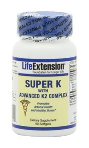 |
| Life Extension Super K with Advanced K2 Complex |
Additional Resources
Cites
- Yadav VK, Ryu JH, Suda N, et al. Lrp5 controls bone formation by inhibiting serotonin synthesis in the duodenum. Cell 2008;135: 825-837.
- GASTROENTEROLOGY 2011;141:439-442
- http://care.diabetesjournals.org/content/34/9/e147.full
- Shea MK, Booth SL, Gundberg CM, Peterson JW, Waddell C, Dawson-Hughes B, Saltzman E: Adulthood obesity is positively associated with adipose tissue concentrations of vitamin K and inversely associated with circulating indicators of vitamin K status in men and women.
- J Nutr. 2010 May;140(5):1029-34
- PMID: 17145139 [PubMed – indexed for MEDLINE]
- Wu S, Liao AP, Xia Y, Li YC, Li JD, Sartor RB, Sun J: Vitamin D Receptor Negatively Regulates Bacterial-Stimulated NF-{kappa}B Activity in Intestine. Am J Pathol. 2010 Jun 21
- http://www.ncbi.nlm.nih.gov/pubmed/16942519
Very interesting read. Especially interested at the moment as I'm taking Warfarin (hopefully for just a few more weeks, then I'll have to find a way to stop taking it) – so I'm drastically low on Vitamin K (I imagine)
Good thing I upped by vitamin K. I have no gallbladder. 🙁 How much do you usually recommend for us maimed ones? 🙂 I'm taking 15 mg.
@colleen. That should be plenty
@Collen:
Where are you able to get such high dosages for a reasonable price? Can you link me?
(I don't have a GB either)
Thanks
Here's what I'm using. I take 3 capsules a day http://www.vitacost.com/Carlson-Vitamin-K2-Menate…
Dr. Kruse,
I wrote about this in another context but I have epilepsy, probably post-traumatic, and have been on phenobarbital therapy for over 40 years. Despite numerous questions to my physicians over the years about the possible side effects of long-term barbiturate therapy, I was never told about it as a potential cause of secondary osteoporosis. I found out after I fractured my pelvis and arm after a minor fall. Of course, the fact that I was 5'11 when I played lacrosse in college and am currently 5'8" should have been another clue.
My attempts to go off phenobarbital and onto another medication have been woefully unsuccessful. My endocrinologist, who specializes in matabolic bone disorders, has me taking high dose Vitamin D2 (50,000 IU 3x weekly). It barely keeps my Vitamin D levels in the 50-60 ng/mL range and I periodically have elevated PTH.
I have started Vitamin K2 (5 mg. daily)and am attempting to follow a ketogenic paleo diet. I'm not overweight, exercise regulary with weights, and my CRP-HS was 0.07 mg/L when last tested in August. Nevertheless, would it help to try a leptin reset?
Or does the phenobarbital (which causes other problems as well, e.g. sex hormones, lipids, etc.)make this a bit hopeless?
@Paul its never hopeless. I think you need a good protein and fat laden diet with 10-20% of carbs. I think supplementing K2 is huge for someone like you because you are on a drug that predisposes to osteoporosis. The next few blogs will address your issues.
At what doses would k2 become a potential problem?
@ CB
Vitamin Research is a great place to buy 15 mg Vitamin K2.
http://www.vrp.com/bone-and-joint/ultra-k2
They have a constantly revolving series of weekly discounts with buy one get one free every couple of weeks.
poor LDL receptor function causing it not to be uptaken quickly enough by the cells of our body. When this occurs we generally also find in serum assays a particle size change to sdLDL and a lowered HDL level.
Well as a Male,my HDL of 59-64 is not low, but i generate lots of particles,probably due to poor LDL receptor. With a family history of CAD I suppose this is the reason. With normal thyroid and D3 in the 40-50 range, any suggestions on how to address poor LDL receptor issues? The CW answer would be statins.
Thanks,
Not sure how you feel about Weston Price, but here is a link to a 2008 article about Vit K, lots of charts, food lists, studies etc… If these views don't jive with yours, please delete this post. http://www.westonaprice.org/fat-soluble-activator…
Dr Kruse,
Could you please mention what are good sources of vitamin K that people can incorporate into their diet and what are optimal levels?
@Jim Yes, bone cells do have nuclear receptors for K2 as well.
@Erin I generally dont treat MS patients but I so think everyone with MS should be tested for an osteocalcin level not so much for their bone but for the myelin protection. K2 is a cofactor in its production in the CNS.
@Selma Vitamin K2 enjoys a very large therapeutic window.
Same K2, much cheaper price (and I'm NOT affiliated with Swanson in any way except as a customer):
http://www.swansonvitamins.com/CSN028/ItemDetail
Darleen's source (300mg total for $16) is considerably more expensive per mg K2 than Adriana G's (1350mg total for $50), not even including the discount for additional bulk purchase for Adriana's.
@Dr. Kruse: Would liver or the heart be good sources of K2 in terms of eating offal?
Is vitamin K2 deficiency the cause of spider veins?
@Darleen – 16×3=$48=more expensive, actually.
@Darleen – 16×3=$48=more expensive, actually.
@Kronk
MK-4 has a very short half-life, about one hour,
it is better to take 3 pills/day and spread them throughout the day.
Or for most, just one pill/day.
.
@owl. No that appears to be related ti estradiol and insulin levels
@akman. I think weston a price was a true giant who got marginalized by the powerful ADA. Im still pised i never learned about him in dental school
Hi Dr. Kruse,
I just went for my bloodwork today. I had a ton of stuff tested and I am anxiously awaiting the results. I did the test fasted, eating nothing from about 8 pm to 6:30 am, but the afternoon before the test i had a centrum multivitamin. in addition to bloodwork, i also am having urinalysis. will that vitamin skew the results of the vitamin level tests i had??
@V the multi could screw up your labs. If any come back really unusual I would consider repeating them if any treatment decisions will be made using them.
ps i read that kurt harris was supposedly making fun of you in emails to other paleo bloggers. right before he banned me from his blog, he implied to me that he and other paleo bloggers were making fun of me (ie the only possible blogger i can think of is dr bg, since i asked her to comment on a question i first asked him). that kind of tactic is very middle school and speaks to him being a major a-hole. Then i read over at PH that you were putting the thread criticizing you over at PH up in your OR. i hope you are not trying to do the same thing as harris- ie ridicule people by letting them know you are your group are laughing at them.
EDIT: No I did it because I needed a laugh. "the management"
anyway, i'm still grateful for any advice/insight you can give me.
everyone needs a laugh. just keep it secret.
I started the l-glutamine for leaky gut this weekend. I have not had any cramps since I started on Friday. I am now taking Vitamin K2 and plan to add back the magnesium malate in about a week. How long should I take l-glutamine to repair my leaky gut, or do I take the l-glutamine for ever? My weight had stabilized at a 15 lb loss until this weekend when I lost another lb. Thanks
Dr. Kruse, I saw you said Vitamin K2 could be helpful with Multiple sclerosis. I have a friend with this disease. any chance I could get you to go into more depth on this, and/or make some more recommendations/suggestions on helpful supplementation? Thanks!
@Steve I will but I want you to look up Dr. Terry Wahls from Iowa. She cured her MS with a paleo diet. https://www.facebook.com/Dr.Terry.Wahls
Her recent TEDx event is a must watch.
Thanks! Actually, I just found her last night from a link on Marks Daily Apple, but glad to see it twice, and a recommendation on her work from another MD.
Great article, Dr. Kruse.
A question: Is it necessary to also supplement with D3 and a source of Vitamin A while supplementing with K2?
If so, do you have a rough ballpark of what a recommended ratio might be?
@Brad you do not need any A if you are on a paleo or primal template in my experience
Dr. Kruse, would K2 supplementation generally be contraindicated in someone with D.I.S.H. (Diffuse Idiopathic Skeletal Hyperstosis) or beneficial? I'm still trying to understand if it is a GLA protein issue and what that means as far as K2. Thanks so much for all you do.
@Kim Im not away of any studies on it. I would not expect it to be a problem at all.
@John B regarding your email on Homocysteine/methylation here is my response.
We convert back to methionine via vitamin B12 and folic acid — part of a process called the methylation cycle.
We then convert into cysteine with the help of vitamin B6 — called transsulfuration process.
Hi Jack
Do you know if K2 can help with bone conditions such as fibrous dysplasia? I have this diagnosed in my femur.
I was a crossfit athlete eating paleo until Nov 2010 when what turned out to be a nerve injury resulting from bone damage caused by FD and my strength/conditioning programme was initially misdiagnosed and instead I was prescribed (for no reason since I had no evidence of an infection) a bunch of drugs – Doxcycline, Ciprofloxacin, an nsaid Diclofenac and then after a near anaphylactic reaction to this lot – diazepam. 13 months later and while signficantly recovered I'm still unable to exercise and live with the remaining ADR's – intra cranial pressure/head aches, tinnitus and fasciculations/peripheral neuropathies and myalgias.
I use green pastures butter oil for K2 and currently eat strict paleo (plus auto-immune protocol no nightshades).
@Geoff I used to see fibrous dysplasia a lot when I was an Oral Surgeon but I have only seen one case since I have been a neurosurgeon and it was in the zygomatic bone and I resected the entire lesion and the skull and rebuilt the lateral half of the face. The guy did very well and had a great cosmetic result. This was a mono ostotic case of the skull and zygomatic root. This disease is associated with elevated levels of the transcription factor C-fos. This leads to gene over-expression and eventual tumor formation. Don't fret because it is not hereditary. I don't know if K2 would have any effect but I will look into it since you asked. It is a very rare disease and I only know about it because oral surgeons see it more frequently than a neurosurgeon. There are generally two types of fibrous dysplasia: 1. Monostotic which involves one bone, or adjacent bones.
2. Polyostotic (Involves several bones not contigous). The most severe form of this disease is polyostotic fibrous dysplasia and is known as McCune-Albright syndrome. I treat one case of this as a resident in OMFS.
Forgot to mention – my orthopedic consultant sent me for endocrinological tests which I was told returned normal. I will check to see if C-fos was assessed. I read that FQ's are known to be involved in abnormal osteoblast differentiation and function. I've also read that FD patients have blood cells with G protein gene mutations (GNAS) and that the mono-stotic variety is normally inactive after puberty. Will do some research on FQ's and GNAS and C-fos to see if any possible connections and continue to eat as primally!
Hi Dr. Kruse,
I have followed the guys and girls to the Leptin reset forums, I need some advice on K2,
I started taking it and it seems that it is making me feel irritable, does it form into glutamine? Sorry if I have this wrong, I have noticed the same day after taking it I feel irritable, almost a sort of anxiety, not sure if it's the K or if it's causing my thyroid medicine to work better and I am feeling those effects.
Glutamine causes me to become aggitated, any advice would be greatly appreciated!
Thank you Dr. Kruse,
Ellie
@Elle K2 does not form into glutamine……ever
Thank you! Do you have any idea why it could be causing aggitation? Perhaps it's helping the amour thyroid work better and I am feeling that feeling, like a hyper sort of feeling? I am going to test this out again on my days off from work, today I was grumpy and I noticed it the other days too, like a too much caffeine type of feeling. The only new thing was vitamin b, k2 and coQ10.
A comment about my skin.
within days my husband looked at my face and said something about how pretty I looked and my son said I was still youthful, it must have brightened up my skin, they had no idea I started taking it. It really made me feel good, so I hope to find the reason for the aggitation. I have been trying to research for the reason, I can't find it.
@Elle my bet is your gut is so leaky it is affecting your ability to make neurotransmitters for your brain and you are feeling the downstream effects of this……..You need to step up your gut protection in my view. Ready my leaky gut Rx stat.
I work in a grocery store and I know I can get bones. Do you know the best bones to ask for? Something else I do think I have a leaky gut problem, I was on antibiotics for a long time in my 20's. I feel this led me to a lot of health problems in my 30's. Thank you!
Ellie
@Ellie any grass fed bones will do. If they are long bones have the butcher cut them for you. Whole foods cuts them for me all the time.
In reference to bone spurs on hands and cervical neck bone spurs. Would K2 be a benefit to reduce these? Or at least, to slow the growth? My mother who is 83 has this and I am most likely genetically inclined.
Dr. Kruse,
I am a 47 year old female and I had my Gall Bladder removed over 21 years ago and I’ve had nothing but stomach problems and cholesterol problems ever since. Recently, I went to see my Chiropractor and took with me x-rays of my lower back and he commented that I had calcium build-up in my arteries. My recent blood test have my HDL at 31, my Triglycerides at 188, LDL at 129. My HDL cholesterol has always been low since my gall bladder problems and have been as low as 24. My vitamin d, 25-hydroxy was 26.7 which is very low. Also, recently I have been diagnosed with MVD (microvascular disease), and these arteries are obviously clogged with calcium and who knows what else. I have been on a vegan diet for the last year due to chest pains and my cardiologist telling me to get my cholesterol down. The vegan diet does not seem to help and I still have chest pains. My question to you is do you think Vitamin K2 could help take the calcium out of my arteries and put it where it belongs? Also, I’m confused as to which Vitamin K2 to take, MK4 or MK7.
Penny
@penny It will help…..mk4 or 7 are fine. I like MK 4 best. Vegan diet is the best way to get heart disease. You need to go 180 to it and eat Paleo. go buy Robb Wolf’s book The Paleo Solution. That is how I eat.
Dr.Kruse, thanks so much for doing what you do! Pioneers and risk takers are my heros. My son introduced me to your site and I will be forever grateful.
I am a 69 yr old female and have been on paleo plan for about 5 months. Have access to grass-fed meat through a buying club. Although never having a weight issue (as an adult), I have lost the waistline ring and clothes fit very differently now. Yea! Best of all I feel great, though never felt badly before. Take no meds; however, was diagnosed with osteopenia in 2001 and warned of osteoporosis. Have never taken osteoporosis prevention meds. Take a multivit/min, (includes about 685 mg calcium citrate and 2000 IU D3) and Omega 3 (502 mg DHA and 1005 mg EPA). Also, have been taking glucosamine/chondrotin for over 10 years after experiencing achy hands at night. This really eliminated the discomfort; do have some arthritis at base of thumbs. Follow Loren Fishman’s yoga exercises for osteoporosis. Just recently added Vit K2 MK4 (about 15 mg/day).
Question: am I on the right track? Haven’t had any labs done. Weight training?
Thanks again!
@joyce You are doing well Read this…..https://jackkruse.com/the-osteoporosis-rx/ I think lifting heavy things and doing paleo are awesome for you…..I also love swimming instead of sprint for my older ladies……it also is built in CT……..
You are awesome! OMG! Forgot to mention losing 4 lower incisors due to periodontal disease last Fall. Got a hard wake up call and a much better dental practitioner. Am working on implants; ouch $$$$. Also, taking Co Q10.
Happy to forego the sprints! One paleo son is a trainer and will help with the weights. Blessings to you!
@joyce with perio disease up your K2 and consider swishing and spitting out coconut oil three times a week…..
Thanks for the update!
Is it OK to take 30mg of K2 together WITH the following:
1000mg Krill oil
1400mg Fish oil
Vit D3-5000
B-complex
Multi-vitamin
Mobic
Omeprazole
Or is spacing these meds better for optimal absorption? Thanks!
Dump the fish oil and up the krill to 1500 mgs……and use spot icing on that hip big time
My krill oil comes in 1000mg tabs. Can I take 2g?
yep…
Hi Jack
I have a slightly high calcium level (10.4) and low PTH (20). Retested 10 times. Some sites would leave one to believe this is hyperparathyroid or cancer. I would like to believe neither. My TSH is 8.5, 25OH D3 27, T3 and T4 free are low normal range. Would k2 make a difference or is there another underlying issue?
@Legend Your TSH is off the charts! Your D is horrible and you’re a wreck!!!! You need help. My bet is you have multiple issues. You need a bio hack….and a good doc.
I forgot to also ask if K2 helps to moderate slightly high RBC HGB and HCT levels. I’ve never taken TRT or EPO and am not an athlete. tks
Hi Jack will K2 be beneficial for a pregnant friend with gestational diabetes? She wants to avoid injecting insulin which her doc is pushing her to do. She also has getstational gingivitis and according to her, her gums “gush blood” when she brushes. I told her to ask her doc about Vitamin K2 before going with the insulin but would be interested in your opinion on K2 during pregnancy. What would be an appropriate dose?
I also linked her to this blogpost so she could show her doc at her appointment on Monday. I told her to make the doctor read it(ipad) while she is in the office with her and to not let the doc give it the brush-off. She says she will do her best.
What about folks who are on coumadin?
@Elizabeth that is when you discuss the situation of moving off coumadin to pradaxa or to lovenox with the doctor.
This site was… how do I say it? Relevant!
! Finally I have found something which helped me.
Kudos!
Hello Doctor,
Because of persistent spine pain at the height of the blades, my doctor told me to do an MRI. As a result, it turned out that I have spine osteophytes there. The doctor told me that I can not take to much calcium because it causes this problem. So my question is if Vitamin K2 may be helpful for me?
Eugene I am no fan of calcium supplements ever. Lack of K2 = lack of UV light exposure over your skin eye gut and lung……so you need to get those surfaces into the sun……..that is the real deficit. You can take K2 but you need to really change your light environment. Read my Ubiquitination 24 blog and make sure you read the Time series of blogs.
Thanks Doc.!
I live in Seattle and there is not to much sunny days. Maybe in the couple of months…
Will K2 help remove Bone Spurs / Calcifications on toes / feet? My diet is superb, avid athlete, take supplements, perfect blood work etc – stumped on how to remove the growths on R foot. (mostly due to being avid athlete w/ slight leg length descrepancy which I’ve now re-corrected w/ a lift) Thanks
Calcium deposits usually come from higher heteroplasmy rates in those areas, lack of sunlight and insulation from both sunlight and the Earth Magnetic flux. Mitochondrial depolarization occurs when calcium rushes into a mitochondria. When this occurs it triggers the Mg2+ release. Calcium is a secondary messenger that takes a light stress response in our eye and alters the RPE in the eye to increase the paraventricular nucleus firing to increase the stress response everywhere and in every mitochondria in cells. This is why calcium efflux from nnEMF and blue light is toxic to cells. Calcium causes mitochondria to swell and change shape. Shape and size changes alter the autophagic and apoptotic programs in mitochondria and this is why calcium is so toxic to mitochondrial size. Blue light expands their size most and UV and IR light shrink their size most. When UV light is not exciting electrons properly in any way, and this is why most neolithic diseases seem to be associated with an insatiable appetite for calcium. This is especially true in cancers. This calcium release stimulates increases the amount of ELF-UV light from fluorophore proteins and respiratory proteins. In cells under stress, they begin to use the IP3/DAG signaling pathways when mitochondrial proteins are expanded on the inner mitochondrial membrane. ELF-UV and cooling both increase catecholamine release from the PVN and this in turn helps increases melatonin secretion in WBC’s to modulate inflammation in the skin and blood plasma. This explains why UV light and cooling are critical aspects of circadian coupling of codons. Codons can code for the same amino acids but certain codons are used with certain light frequency and when temperatures are also linked to this process. This is how the cell controls different dopamine receptors in summer time and different ones in winter time. This is why a Warburg metabolism is used as signal of altered light frequencies at some level in the system because all the aromatic amino acids capture sunlight. And all aromatic amino acids make biogenic amines with UV light and cold. The key is realizing different codons are used in different seasons to build proteins. The aromatic amino acids interaction with ELF-UV light release from cells are what makes things like the biogenic amines from ELF-UV function physiologically differently even though they may look identical at a protein level. They are not. Every biogenic amine is made this way and in a cancer state/neurodegeneration state, there is fewer biogenic amines being made in high enough quantities to gt proper functioning. This is why aromatic amino acids are up-regulated in a Warburg metabolism. It is also why glutamine is up-regualted too.
In stressed cells, from too much exercise or in cancer cells, and most illness states there are always abnormal hormone panels. Why? What control pituitary hormone release? Dopamine and prolactin. Both of them are biogenic amines linked to these aromatic amino acid codons. Solar light builds our hormone panels by building dopamine in our eye, retina, and frontal lobes using aromatic amino acids that absorb UV light. Athletes with calcium deposition usually have trashed hormone panels because they have a life filled with circadian mismatches.
Vitamin K2 is so important for the body.
Hi is 15000 mcg vitamin k safe ? Does k1 interfere with k2 absorption ? I read that k1 can increase k2 levels higher than k2 is this true ? Doe’s mk4 and mk7 compete for absorption ? Thank you.
You can contact a pharamcist or http://www.lef.org to get this answer.